3D anatomical atlas that provides detailed information about brain regions, connectivity, and function, to support neuroanatomy research.
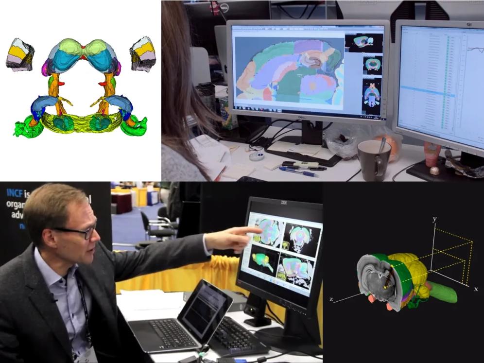
- Explore detailed 3D delineations of brain regions in an intuitive web-based atlas viewer.
- Find your brain region of interest easily with the linked brain region hierarchy.
- Find and access data shared through EBRAINS and registered to the Waxholm Space Rat Brain Atlas.
- Use the atlas embedded in other EBRAINS tools, allowing data integration and automated spatially linked image analysis.
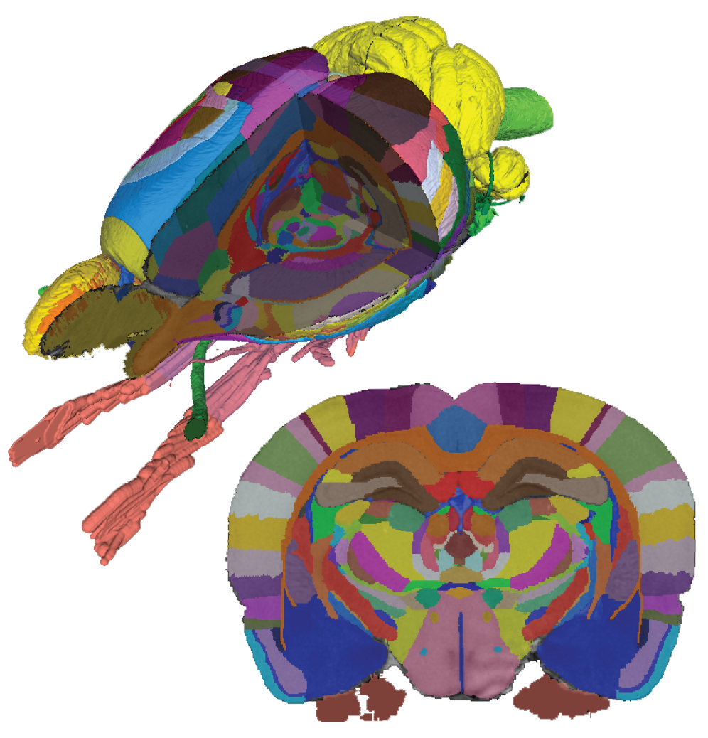
The Waxholm Space rat brain atlas is a detailed volumetric atlas of the rat brain, to which a wide range of anatomical and functional data have been registered, including detailed data showing cellular distributions, axonal pathways, and gene expression patterns. EBRAINS provides a visualization interface, enabling researchers to explore and compare different aspects of the rat brain in 3D space.
Explore or download the Waxholm Space Atlas of the Sprague Dawley Rat Brain
The Waxholm Space (WHS) Rat Brain Atlas contains comprehensive anatomical delineations of the adult rat brain's brain regions and fibre tracts. The atlas was acquired from an 80-day-old male Sprague Dawley rat using high-resolution ex vivo magnetic resonance and diffusion tensor imaging (MRI/DTI) at the Duke Center for In Vivo Microscopy (Durham, NC, USA). It includes spatial coordinates and the position of bregma and lambda, facilitating conversion to stereotaxic coordinates.
The atlas, the MRI/DTI volumes, and the diffusion tensor data are available, along with labels and configuration files for ITK-SNAP, the Mouse BIRN Atlasing Toolkit, and PMOD. The atlas is shared in a standard volumetric format (NIfTI) containing delineations of 222 brain regions, accompanied by a label file with the name and ID of each region.
Waxholm Space Rat Brain Atlas from NITRC
Download
Atlas dataset and publications
Find datasets
Related tools
The Waxholm Space Rat Brain Atlas is the backbone of spatially-focused workflows and tools that support spatial registration of new data to the atlas template, semi-automatic analyses using the brain region hierarchy and delineations, and visualisation of extracted data in 3D.
The atlas is incorporated in the QuickNII tool for spatial registration of serial 2D images, and employed in the QUINT workflow for extracting and quantifying labelled objects from images registered to the atlases.
Learn more about the QuickNII tool
Learn more about the QUINT workflow
Connect to atlas developers and the user community
The Waxholm Space rat brain atlas is in active development by the atlasing team at the Neural Systems Laboratory, Institute of Basic Medical Sciences, University of Oslo, with contributions from experts on the anatomy of individual brain regions. The level of detail in the atlas is evolving, with each subsequent version offering new and refined anatomical delineations.
Downloads
As of October 2023, the WHS rat brain atlas has been downloaded 33000 times.
Contributors
Over 20 anatomists, experimental neuroscientists, students of medicine and neuroscience, software developers.
The atlas repository at the NeuroImaging Tools & Resources Collaboratory (NITRC) is accompanied by a public forum that gives you the opportunity to ask questions, suggest improvements, and contribute to the discussion around the use and development of the atlas. A brief introduction to the latest developments is given in the atlas wiki.
The anatomical delineations, the MRI template, as well as surface representations of each anatomical structure have also been incorporated into the Analysis of Functional NeuroImages (AFNI) toolbox. For AFNI-specific issues, please refer to the AFNI message board.
Join the discussion on NITRC
User forum
See the latest developments
Wiki
Learn more about using the atlas in AFNI
Message board